Exercise 3A: Cell Replication
Purpose
In this lab, we were investigating the process of mitosis by looking at slides of onion root tip. We were looking at the slides in order to see the products as well as the relative duration of the different phases throughout mitosis. By viewing the onion root tip cells, we were able to identify the different stages of mitosis in plant cells.
Introduction
In eukaryotes, mitosis is the process in which cell division results in two daughter cells with identical sets of chromosomes. The first and longest stage of mitosis is known as prophase, in which chromosomes become visible and centrioles separate in order to move to opposite poles of a cell. The second stage is called metaphase and this is where the chromosomes line up in the middle of the cell and connect to the spindle fibers at their centromere. The third step is anaphase, where the sister chromatids separate into individual chromosomes and they pull apart from each other. The fourth step is telophase and this is the last stage where the chromosomes all gather at opposite sides of the cell and they lose their rod like shapes. New nuclear membranes form around each DNA and the spindle fibers disappear. Cytokinesis occurs after the fourth step and the cell membrane pinches so that the cell finally divides into two.

Methods

The above picture illustrates a root cell during Interphase, or, the time during which the cell is not dividing. While the nucleus of the cell is visible (the red dot in the center). There is no chromatin evident under the lens of the microscope.

Now the cell enters the first phase of Mitosis, better known as Prophase. During Prophase, the cell begins to condense its chromosomes in preparation for division. If you look closely above, you may see small dots within the nucleus. The chromosomes are now visible simply because they are so concentrated within the nucleus of the cell.

This picture shows the cell in Metaphase, which is identiifed by the chromosomes lining up along the center of the cell.
In this picture, we see two nuclei and a cell plate, which means the cell is in Telophase. Once the cleavage has formed, cytokinesis will officially divide the cell into two identical daughter cells.
Data

This table shows the amount of each phase within each field. The time in each stage was found by multiplying the percent of total cells counted x 1,440 minutes. Interphase had the longest time and Anaphase had the least.
Graphs and Charts

This is a pie chart of the onion root tip cell cycle and the data was provided by the table above!
Discussion
Conclusion
This lab served as an effective means to show real cells in each stage of mitosis. While it is good to describe each stage of the cell cycle, being able to recognize what process a cell is undergoing in real life is still a valuable skill. Aside from allowing us to see plant cells up and close, this is a powerful tool for reviewing Mitosis.
References
http://www.cellsalive.com/mitosis.htm
http://www.marietta.edu/~biol/introlab/Onion%20root%20mitosis.pdf
Exercise 3B: Meiosis
Purpose
In this experiment, we were simulating the stages that occur in meiosis, a type of cell division in sexually reproducing eukaryotes, with the use of chromosome models. With the use of these models, we were able to look at crossing over, when homologous chromosomes exchange parts, as well as the recombination that occurs in the process of meiosis.
Introduction
Meiosis is the shuffling process of genes that occurs when organisms such as plants, animals, and sometimes fungi are ready to reproduce. Meiosis begins in prophase I; meiosis is when two diploid cells divide and then divide again resulting in four haploid cells. Genetic variation through meiosis occurs when maternal and paternal chromosomes cross over and exchange their segments. In metaphase I, the tetrads line up and in anaphase I the pairs of homologous chromosomes split up.
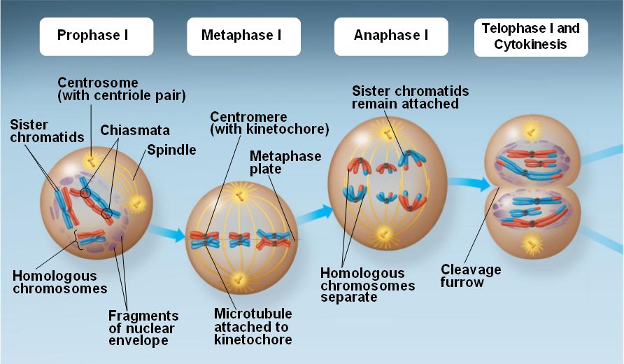
The summarized steps of Meiosis
Methods/Discussion


In Prophase I, the chromatin begins to condense and prepare for cellular division


The final result of "Crossing Over" One red bean and one yellow bead are exchanged to make the gametes different from their parents.

The area where Crossing Over ocurred is where the chromosomes will line up along the center of the cell.

Now the Sister Chromosomes separate, with one chromosome going to one side of the cell and part of it moving with the other chromosome to the other side of the cell.

The chromosomes then move to opposite ends of the cell and a nuclear envelope begins to form to separate the two chromosomes. Two haploid cells are thus formed and then the organism moves onto Interphase II (AKA interkinesis).

These next pictures show what happens during Meiosis II where the chromosomes are slip in order to produce two more daughter cells. In this phase (Prophase II) centrioles move to the opposite sides of the cell.

The chromosomes then move to the middle of each of their own cell.

The centromeres and chromatids separate and move to the opposite sides of the cell.

Finally the chromosomes are all separated and begin to form their own nuclear envelopes. The result is four daughter cells.
Data

This table shows the similarities and differences of mitosis and meiosis.
Discussion
Discussion
Conclusion
The use of beads in this lab provides an effective way to view chromosomes conceptually. By using two colors (red and yellow) we are able to distinguish one chromosome from another and see how the process of crossing over occurrs and how this leads to genetic variability among gametes. We can look at a cell to understand Meiosis, but we cannot know how the cell undergoes this process without focusing on the chromosomes within.
References
http://www.marietta.edu/~biol/introlab/Onion%20root%20mitosis.pdf
http://www.biology4kids.com/files/cell2_meiosis.html